|
|
|
|
|
 | ||
|
| ||
 October 13, 2004
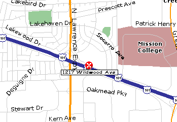
Ramada Inn
PLEASE RESERVE IN ADVANCE --
The Micro-XCT includes a fully automated tomographic data acquisition system and real-time reconstruction engine. An interactive 3D visualization package allows the user to view and analyze the 3D data in various ways, including 3D rendering and virtual cross-sectioning and de-layering. The system's unique capability of imaging at high-resolution without compromising throughput makes it a powerful tool in non-destructive imaging applications for Advanced Package FA as well as microtechnology and biotechnology (for example, imaging embedded MEMs structures, material stress failure mode analysis, bone implant interface, etc). Since it is non-destructive it is ideally suited for stress and thermal fatigue testing, new process development and failure analysis.
"µCT Techniques In Combined 2D/3D High Resolution X-ray Systems" -- All physical objects, we know, are 3-dimensional. But X-ray inspection shows only 2-dimensional shadow images of their absorption structure. Micro- or even nanofocus X-ray systems provide 2D resolution below 1 µm for inspecting small hidden structures - widely used in the electronics industry (BGA, Flip Chip, CSP, bond wires, die-attach, PCB, etc.) For many applications images of 2D X-ray inspection provide sufficient information for conceiving even the third dimension of a sample. Nevertheless, recent developments have made available various techniques for complete 3D reconstruction of small samples using axial or planar computed tomography. The talk will give an overview on that technique, showing practical examples and images.
Udo E. Frank is the Director of Technology Development of FEINFOCUS GmbH, Germany. He studied physics and chemistry, and received his Ph.D. in molecular physics from the University of Ulm, Germany. Dr. Frank joined FEINFOCUS in 1992, and has gained a comprehensive knowledge of high-resolution X-ray microscopy. He is an expert especially in the inspection of electronic components. During the last five years, under his direction, the FEINFOCUS R&D team has developed several innovative high-resolution X-ray systems with revolutionary new operating and functional designs, several of which have been honored with respected industry awards.
|
SCV Chapter
Home Page |
How to Join IEEE |
Contact our Chapter Chair |
| CPMT Society
Home Page |
IEEE Home Page |
Email
to Webmaster |
Last updated on